


COKSARROSIS- This is arthrosis of the hip joint.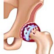 It develops gradually, for several years, prone to progression, can be a double side.It is accompanied by pain and restriction of movements in the joint.In later stages, atrophy of the hip muscles and limb shortening are observed.The diagnosis is established based on clinical symptoms and radiography results.In the early stages of coxartrosis, conservative treatment.With the destruction of the joint, especially in patients with young and a half age, surgery (endoprostrate) is indicated.
general information
COKSARTROSIS (osteoarthritis or deformed hip joint arthrosis) is a degenerative-distribution disease.Usually develops at 40 or older.It can be the result of various lesions and joint diseases.Sometimes it occurs for no apparent reason.COKSARTROSIS is characterized by a gradual progressive course.In the early stages, conservative treatment methods are used.In later stages, the joint function can only be restored operational.
In orthopedics and traumatology, coxarthrosis is one of the most common arthrosis.The high frequency of its development is due to a significant load in the hip joint and the widespread prevalence of congenital pathology - joint dysplasia.Women suffer from cooksartrose a little more often than men.
The CAKSARTROSIS CAUSES
Primary (unknown reasons) and secondary (developed as a result of other diseases) are distinguished.
Secondary COKSARROSIS may be the result of the following diseases:
- Hip articulation dysplasia.
- Innate laxing of the thigh.
- Permanent diseases.
- Aseptic necrosis of the thigh head.
- Infectious lesions and inflammatory processes (eg hip joint arthritis).
- Injuries (traumatic displacements, hip neck fractures, pelvic fractures).
COKSARTROSIS can be one or double.With primary thigh, a concomitant lesion of the spine (osteochondros) and the knee joint (gonarthrosis) is observed.
Risk factors
Among the factors that increase the likelihood of the development of coxarthrosis include:
- Increased constant load in the joint.More often observed in athletes in people with overweight.
- Circulatory disorders, hormonal changes, metabolic disorders.
- Spine pathology (kyphosis, scoliosis) or stop (flat feet).
- Elderly and senile age.
- A sedentary lifestyle.
COKSARTROSIS itself is not inherited.However, certain characteristics (metabolic disorders, structural characteristics of the skeleton, and cartilage weakness) can be inherited by the parents' child.Therefore, in the presence of blood relatives suffering from thigh, the probability of occurrence of the disease is slightly increased.
Patanatomy
The hip joint is formed by two bones: ileum and femoral.The thigh head is articulated with the iliac bone acetabulum, forming a peculiar "hinge".During the movements, the acetabulum remains motionless and the femoral head moves in various directions, ensuring flexion, extension, kidnapping, bringing and rotational hips.
During the movements, the joint surfaces of bone without obstacles to each other, thanks to the soft, elastic and durable Hyalin cartilage, covering the cavity of the revolving cavity and the thigh head.In addition, hyaline cartilage performs a shock absorption function and is involved in the redistribution of the load during motion and walking.
In the joint cavity, there is a small amount of joint fluid, which plays the role of lubrication and provides nutrition of hyaline cartilage.The articulation is surrounded by a dense and strong capsule.Above the capsule, there are large femoral and glute muscles, which provide joint movements and, together with hyaline cartilage, are also shock absorbers who protect the articulation against injuries with unsuccessful movements.
With thigh, joint liquid becomes thicker and more viscous.The surface of dry hyaline cartilage loses its softness, covered with cracks.Due to the roughness that emerged, the cartilage during movements is constantly injured by each other, which causes its thinning and aggravates pathological changes in the joint.As coxarthrosis progresses, bones begin to deform, "adapting" to increased pressure.Metabolism in the joint is deteriorating.In the posterior stages of coxartrosis, severe atrophy of the painful limb muscles is observed.
Caxartrosis symptoms
The main symptoms of the disease include pain in the joint, inguinal, thigh and knee joint.In addition, with ckesarthrosis, rigidity of movements and rigidity of the joint, march disturbance, lameness, hip muscle atrophy and limb shortening next to the lesion.A characteristic of cOKSARROSIS is a kidnapping restriction (for example, the patient is difficult when trying to sit in a chair).The presence of certain signs and their severity depends on the stage of coxartrosis.The first and most constant symptom is pain.
Node1st degree cOKSARROSISPatients complain of periodic pain, which occurs after physical activity (running or prolonged walk).The pain is located in the joint, with less frequency in the thigh or knee.After rest, it usually disappears.The march for 1st degree thigh is not broken, the movements are preserved in full, there is no muscle atrophy.
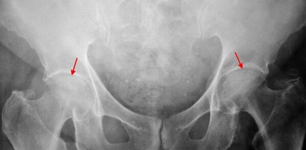
About the X -ray of the patient who suffers from 1st degree thigh, light changes are certain: moderate unequal narrowing of joint gap, as well as bone growths around the outer or internal border of the acetabulum in the absence of head and neck changes of the femur.
NodeCOKSARROSIS 2 degreesThe pain becomes more intense, usually appears at rest, radiating in the thigh and groin.After significant physical activity, the patient with cOKSARTROSIS begins to bear.The volume of joint movements decreases: the kidnapping and the inner thigh rotation are limited.
In the X -ray images for 2nd degree coxartrosis, a significant unequal narrowing of the joint gap is determined (more than half of the normal height) is determined.The femoral head is slightly moved upward, deformed and increases the size, and its contours become irregular.Bone growths with this degree of coxarthrosis appear not only in the internal, but also on the outer edge of the acetabulum and leaves the cartilage.
NodeCOKSARROSIS 3 degreesThe pain becomes constant, worrying about patients not only during the day but also at night.Walking is difficult when moving, a patient with cOKSARROSIS is forced to use a cane.The volume of moving in the joint is markedly limited, the buttock muscles, hips and fighter legs are stunted.The weakness of the muscles removed from the thigh becomes the cause of the pelvis diversion on the front plane and shortening the limb on the sore side.To compensate for shortening, a patient who suffers from cOKSARROSIS by walking leans the body to the painful direction.For this reason, the center of gravity changes, the load in the painful joint increases markedly.
About Third Degree Coxarthrosis radiographs, a marked narrowing of the joint gap, a pronounced expansion of the thigh head and several bone growths are detected.
Diagnosis
The diagnosis of coxarthrosis is based on clinical signs and data from additional studies, whose main radiography is.In many cases, X -rays make it possible to establish not only the degree of thigh, but also the cause of their occurrence.Thus, for example, an increase in neck diaphysal angle, scenes and acetabulus flattening indicate dysplasia, and changes in the form of the proximal part of the femur are indicated that cOKSARROSIS is a consequence of permanent disease or juvenile episolise.In radiographs of patients with thigh, changes may also be detected indicating lesions.
Like other instrumental diagnostic methods of coxarthrosis, CT and magnetic resonance imaging can be used.Computed tomography allows you to study pathological changes in detail by bone structures in detail, and magnetic resonance imaging offers the opportunity to evaluate soft tissue disorders.
Differential diagnosis
First of all, coxarthrosis should be different from gonarthrosis (knee joint osteoarthritis) and spine osteochondrosis.Muscle atrophy, which occurs in 2 and 3 stages of thigh, can cause knee joint pain, which is usually expressed brighter than pain in the area of damage.Therefore, with the patient's complaints about knee pain, a clinic (inspection, palpation, determination of movement volume) is the study of the hip joint and, if there is suspected coxarthrosis, to direct the patient to radiography.
Pain for root syndrome (nerve root compression) for osteochondrosis and some other spinal diseases may imitate pain with thigh.Unlike cOKSARTROSIS, when tightening the roots, the pain occurs suddenly, after unsuccessful movement, a sharp curve, lifting weights, etc., is located in the buttock area and spreads along the back of the thigh.A positive symptom of voltage is detected - intense pain when the patient tries to lift a straightened limb, lying on his back.At the same time, the patient freely takes his leg to the side, while in patients with cksartrosis, the kidnapping is limited.It should be borne in mind that osteochondrosis and cOKSARROSIS can be observed at the same time, so in all cases, a thorough examination of the patient is required.
In addition, cokeArtrosis is differentiated to exchange (boot burrs) - aseptic inflammation in the area of glute muscles fixation.Unlike thigh, the disease develops rapidly within 1-2 weeks, usually after a significant injury or physical activity.The intensity of pain is greater than with cOKSARTROSIS.Movement limitations and limb shortening are not observed.
In some cases, with an atypical course of the disease or reactive arthritis, symptoms may be observed that resemble thigh.Unlike thigh, with these diseases, the peak of pain falls into the night.Pain syndrome is very intense, can decrease when walking.Morning stiffness is characteristic, which occurs immediately after waking up and gradually disappears within hours.
COXARTROSIS TREATMENT
Pathology treatment is involved in traumatologists orthopedists.The choice of treatment methods depends on the symptoms and stage of the disease.In 1 and 2 stages of coxarthrosis, conservative therapy is performed.During the period of coxarthrosis exacerbation, injection blocks, non -esteroid anti -inflammatory drugs (pyroxes, indomethacin, diclofen, ibuprofen etc.) are used.It should be borne in mind that the medicines in this group are not recommended for a long time as they may have a negative effect on the internal organs and suppress the capacity of the restoration hyaline cartilage.
To restore damaged COKSARTROSIS cartilage, funds from a chondroprotective group (chondroitin sulfate, cartilage extract etc.) are used.To improve blood circulation and eliminate spasms from small vessels, vasodilating medications (zinnarisine, nicotine acid, pentoxifiline, xanthinol nicotinate) are prescribed.According to indications, muscle relaxants (muscle relaxation drugs) are used.
With stubborn pain syndrome, patients suffering from cOKSARROSIS can be prescribed intra -articular injections using hormonal medications (hydrocortisone, trimcinolone, metrumor).Steroid treatment should be performed with caution.In addition, with thigh, local products are used - hot ointments that do not have a pronounced therapeutic effect, however, in some cases they relieve muscle spasm and reduce pain due to their "distracted" action.In addition, with thigh, physiotherapeutic procedures (ultrasonic therapy, laser treatment, UHF, inductothermia, magnetotherapy), massage, manual therapy and therapeutic gymnastics are prescribed.
COKSARTROSIS Diet has no independent therapeutic effect and is used only as a means of reducing weight.Reduction of body weight allows you to reduce the load in the hip joints and, as a result, facilitate the course of cOKSARTROSIS.To reduce the load in the joint, the doctor, depending on the degree of thigh, may recommend walking with a sugarcane or crutches.
In the posterior stages (with third degree thigh), the only effective treatment method is the operation - replacing the joint destroyed with an endoprhesis.Depending on the nature of the lesion, it is a single unique pole (replacing only the thigh head) or two -piles (replacing the thigh head and the rotating cavity) can be used.
The endoprothetic operation for coxarthrosis is performed in a planned manner after a complete examination under general anesthesia.In the post -operative period, antibiotic therapy is performed.The seams are removed within 10 to 12 days, after which the patient is prescribed for outpatient treatment.After endoprostrate, rehabilitation measures are necessarily performed.
In 95% of cases, surgical intervention to replace coxarthrosis joint ensures a complete restoration of limb function.Patients can work, move actively and even play sports.The average life of the prosthesis, subject to all recommendations, is 15 to 20 years.After that, a second operation is required to replace a spent endoprosthesis.